Pediatric cardiologist Piers Barker, MD, never forgets a heart, and especially not a rare one. That’s why he was taken aback when flipping through cardiac images for a new six-year-old patient he was scheduled to see at Duke Children’s Hospital in 2006. He was sure that he had seen the heart before, but he didn’t recognize the patient’s name.
It turns out that he had seen the heart of Josie Dunnigan before, when he was a pediatric cardiology fellow at the University of Michigan helping care for an infant who had just undergone a very complex heart surgery. Her name back then was Josie Plum, and she was born with a very rare condition called congenitally corrected transposition of the great arteries. When this happens, explained Barker, the ventricles, or pumping chambers the heart, are on the opposite side compared to normal.
“Josie’s left ventricle which usually pumps blood to the body, was on the right side of her heart, and the right ventricle, which usually pumps blood to the lungs, was on the left side of her heart,” said Barker, a professor of pediatrics in the School of Medicine. The most severe instances of the condition occur in about 1 in 25,000 live births.
Josie became one of the first children in the United States to have what’s known as a double-switch procedure to re-direct blood flow and re-assign the left ventricle with the task of pumping blood out to the body, as it should. She also received a new pulmonary valve to restore normal blood flow to the lungs.
Since her first heart surgery in 2001 at the age of one and a half, Josie and her family had moved to North Carolina and her mother sought out Dr. Barker for her daughter’s continued care. From 2006 onward, Dr. Barker has served as Josie’s primary cardiologist, seeing her through a total of six heart surgeries by the age of 21.
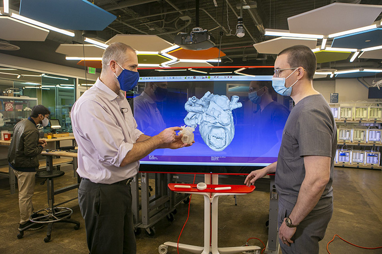
Although she had gone for about eight years without needing a new surgery, last year, Josie’s care team identified a pulmonary valve that needed to be replaced. Considering that the valve was in a hard-to-reach spot in Josie’s unusual heart, Barker determined that she would be a good candidate for 3D heart printing, a unique collaboration between the Duke Children’s Hospital and the Duke Innovation Co-Lab, an on-campus creativity incubator.
With detailed imaging data, the Co-Lab was able to print a 3D model of Josie’s heart and ribcage to help the surgeons plan the procedure.
“The partnership that Duke’s medical center has with the 3D printing lab on campus is really remarkable,” said Barker. “3D printing really fulfills the ultimate goal of non-invasive imaging because we actually have the patient's heart — to scale — in our hands and we can look at it, turn it around, and remove different segments. Similar to a dollhouse, we can look inside and see how all the different sections relate. We can really understand the intracardiac anatomy in a way that is almost impossible to do with two-dimensional imaging techniques.”
The 3D printing program we have here at Duke is really remarkable. It highlights the strengths we have by being a medical center physically co-located with the larger university and the strengths that those two pieces can bring together when they work together.
Dr. Piers Barker, Pediatric Cardiologist
A Heart You Can Hold in Your Hands
Creating a 3D printed heart for a patient is a team effort. First, Greg Sturgeon, a sonographer working with Barker in the Duke Children’s Hospital, was able to take images from Josie’s CT scan and craft them into 3D surface models of her heart and rib cage that he then sent to the co-lab. There, co-lab director Chip Bobbert sent the surface models to the state-of-the-art Stratasys J750 Digital Anatomy 3D printer which is loaded with liquid resin. Over the course of more than twelve hours, the mechanical arms inside the printer spread the resin out in thin, sediment-like layers. A light is then applied to harden the model into a pliable life-sized, perfect replica of Josie’s heart and rib cage.
Josie’s surgery team, Barker, Joe Turek, MD, PhD, associate professor of surgery and pediatrics and chief of pediatric cardiac surgery at Duke Children’s Hospital, and Gregory Fleming, MD, associate professor of pediatrics and director of interventional catheterization, then used the model to come up with a unique plan to replace the pulmonary valve.
Rather than opening up the chest cavity or inserting a catheter through the groin — typical procedures that were not desirable in Josie’s case — the team decided they could make an incision in her side and reach through the rib cage to get to the valve.
Before 3D printing, cardiac surgeons had to rely on viewing a two-dimensional echocardiogram or CT scan and then hold the image in their minds in order to envision the procedure, mused Turek. “But when you are actually able to hold it in your hand, and you’re able to really plan out the operation to a tee, patients are going to have less time on the operating table and therefore will move through hospitalization much faster,” he said.
Paying It Forward
Josie’s surgery on December 10, 2020 went remarkably well, and Josie has been able to continue all of her favorite activities such as dancing, spending time outdoors, and hanging out with her family. Despite having a heart condition and having to go through so many surgeries, having a healthy, active life is something that’s really important to her, and it’s a goal that Barker has also supported as her doctor.
“When patients get treated for things that are not so minor, sometimes they're worried about not being seen as a person, just being seen as their condition,” said Josie. “But with Dr. Barker, I really feel like he cares about me as a person. He doesn't just want me to be successful from a medical standpoint, he wants me to be able to do whatever I want to do in my own life outside of this, and that really makes a difference.”
The excellent and compassionate care that Josie has received at Duke has led her to pursue a career in healthcare. She is training to become an EKG technician at Wake Technical Community College, learning to administer the electrocardiograms that she received so often growing up. Josie said she has been inspired by Dr. Barker, and also by a woman she knew as ‘Miss Mary’ who did her own electrocardiograms from the time Josie was six until she was 17, when Miss Mary retired.
She always had this big bucket of stickers, and it didn't matter if I was six, or if I was 17. She was always like, ‘grab some stickers before you leave’!” said Josie, laughing and then growing thoughtfully quiet. “A lot of times in the medical field, people think you have to be a doctor to make an impact, but having been a patient for so long, you really start to notice that it's sometimes those people in between that really make a big impact in your life. I just really like the idea of being able to be that positive influence for a child that has to come the hospital.”
During a follow-up visit with Barker, Josie got to see the 3D print of her heart and rib cage that guided her most recent surgery. She thought it was “cool” and “bigger than I thought.” She also got to meet Greg Sturgeon, the sonographer responsible for providing the precise 3D surface models of her heart that made the printing possible.
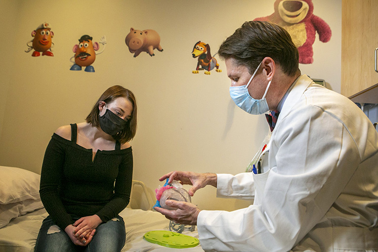
“It was very meaningful to meet Josie and perform her echo, in a follow-up clinic appointment. I was so familiar with her 3D model, it was amazing to meet her as a person and see her unique heart in real time on the screen,” said Sturgeon. “It’s a privilege to be part of her care and to provide these heart models to the surgeons.”
The Co-Lab’s Chip Bobbert is also grateful to have been part of caring for such a special patient at Duke.
“I think I can safely say that pretty much everybody that works in technology hopes that the technology that they're providing, the stuff that they're designing and engineering, is going to improve the world and leave it a better place,” Bobbert said. “Being able to support the medical staff in any kind of way, even if it's in a small way, is just an awesome feeling.”
This article originally appeared on Duke Stories.